
 | Case 23-01: Soft Tissue |
7-year-old female with a right medial knee mass.
Author & Contributor: Bradford J. Siegele, MD, JD, Children’s Hospital Colorado
Case History and Virtual Slide:
A 7 year-old female with no significant past medical history presented to an outside hospital with swelling and possible associated infection versus abscess formation after an episode of blunt trauma to the right medial knee. The swelling resolved without antibiotics, however, nine months later the patient’s mother observed a soft tissue mass in the same site that did not resolve with a course of antibiotics. Magnetic resonance imaging (MRI) showed a “circumscribed multicystic lesion containing fluid levels with thin peripheral and septal enhancement within subcutaneous soft tissues,” most concerning for a lymphatic malformation.
An excisional biopsy of the lesion was performed, with a post-operative MRI showing a residual “5 mm ovoid T2 hyperintense, T1 hypointense enhancing focus within the medial knee subcutaneous soft tissues.” (Fig. 1). Repeat excision was subsequently completed due to concern for residual disease.
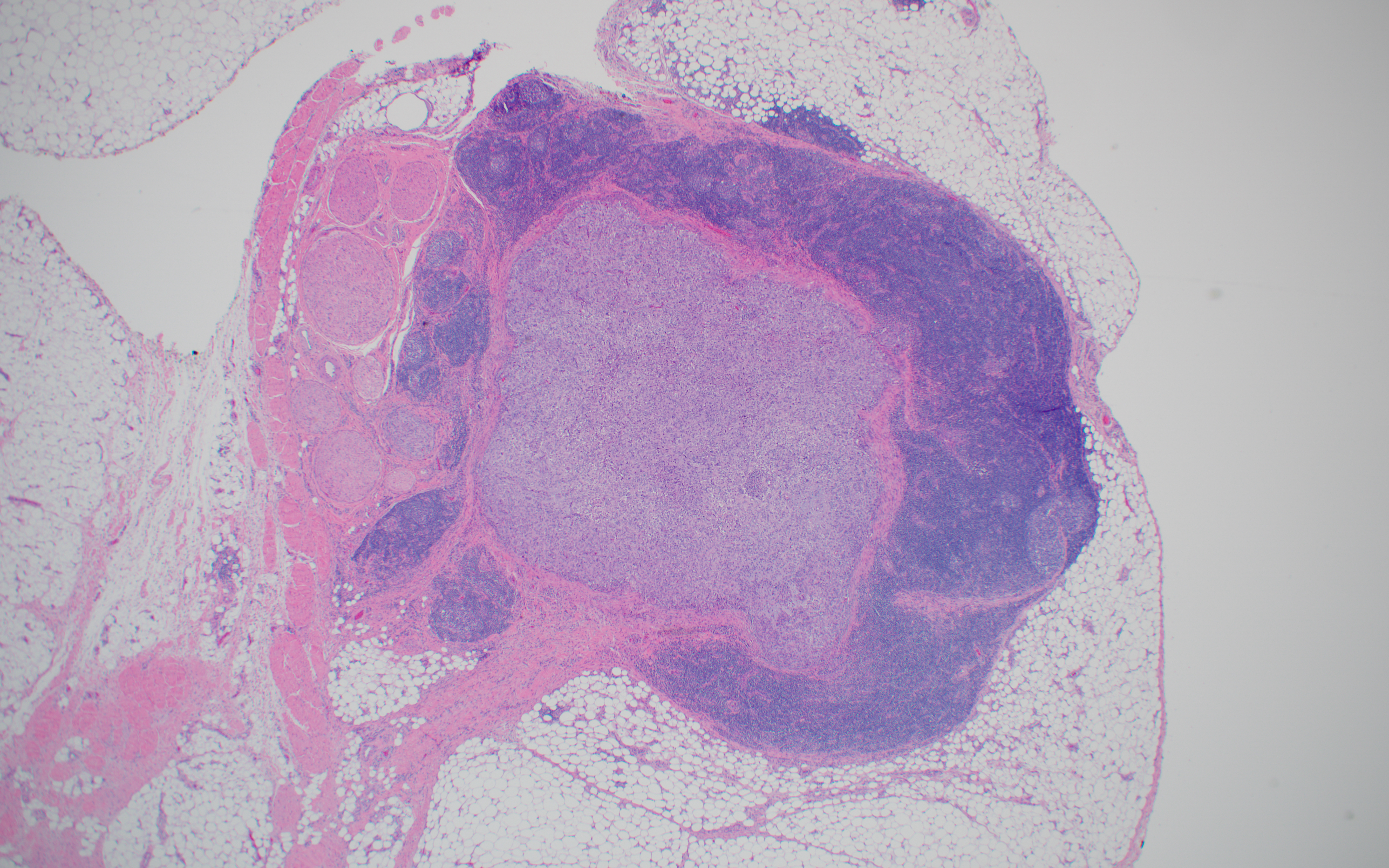
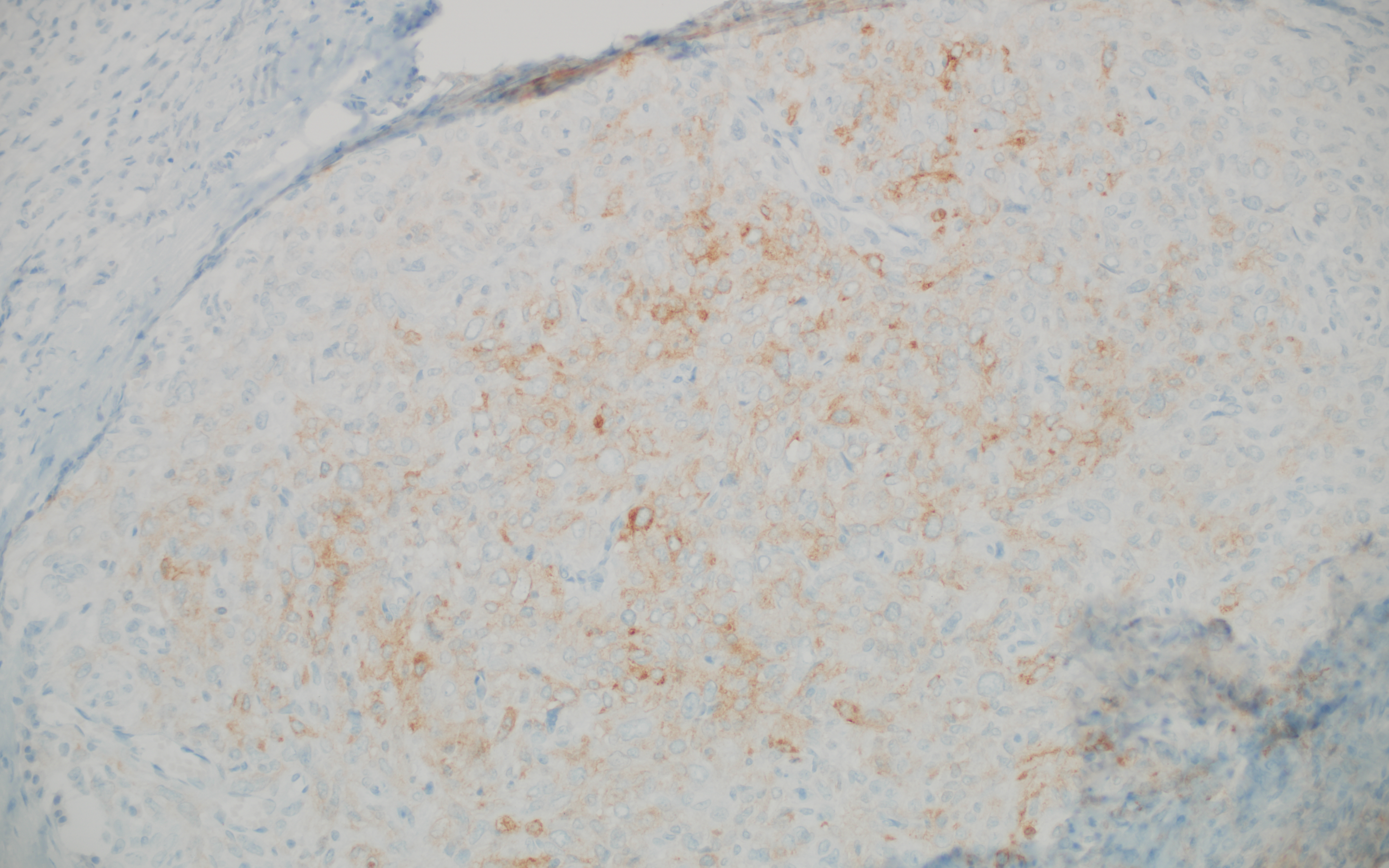
A whole slide image (H&E) of representative tissue from the repeat excision is provided (see also Figs 2-4), as are immunohistochemical stains for CD99 (Fig. 5) and ALK1 (Fig. 6). Desmin and EMA (not shown) were positive. Conventional cytogenetics identified a t(2;22)(q33;q12) translocation.
Access Slide Viewer Here - Slide 1
Access Slide Viewer Here - Slide 2
Fig. 1 | |
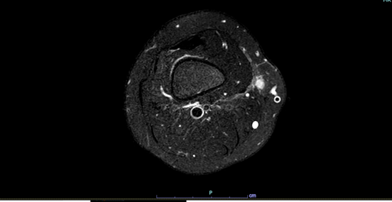 | |
Fig. 2 | |
| |
Fig. 3 | |
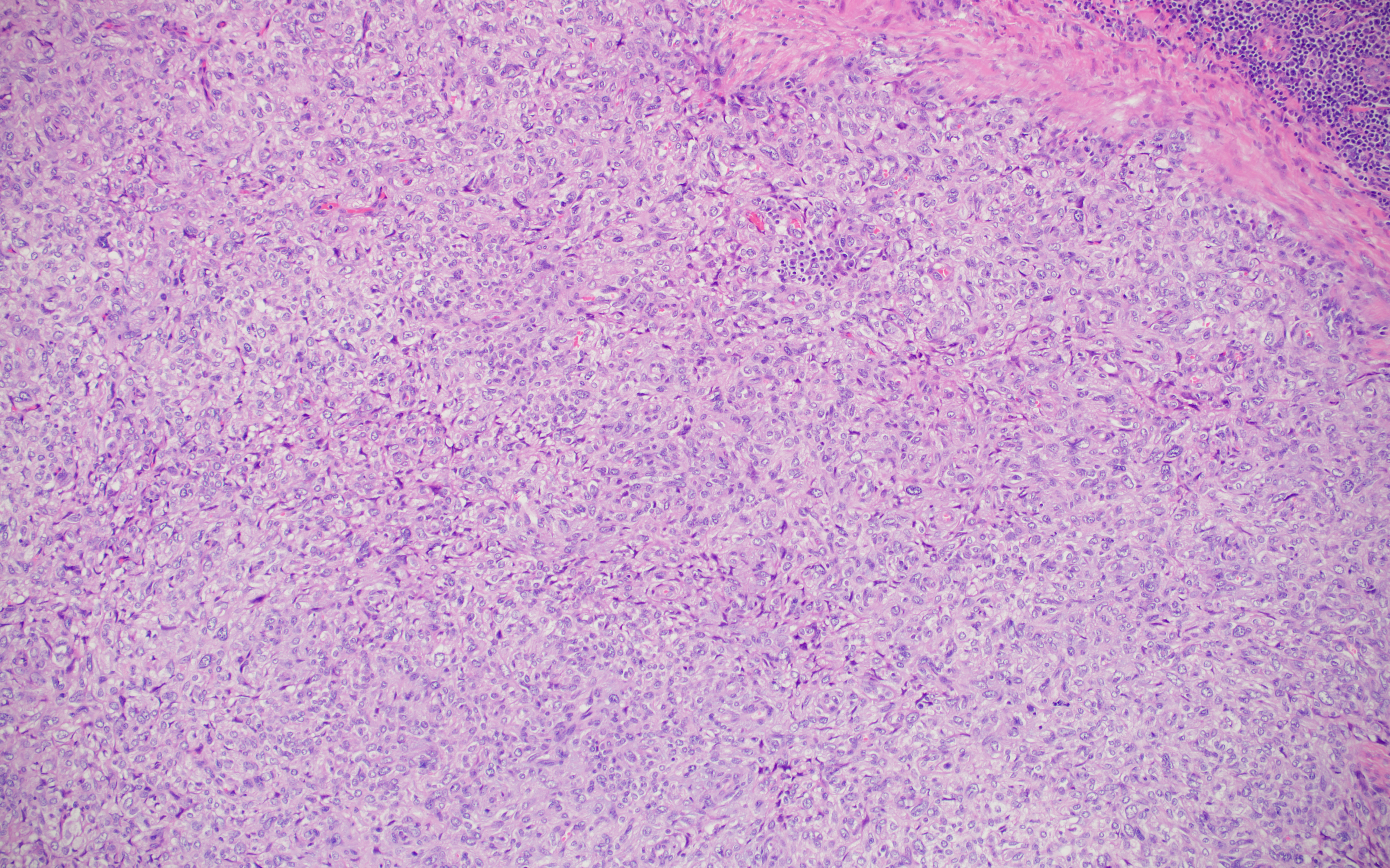 | |
Fig. 4 | |
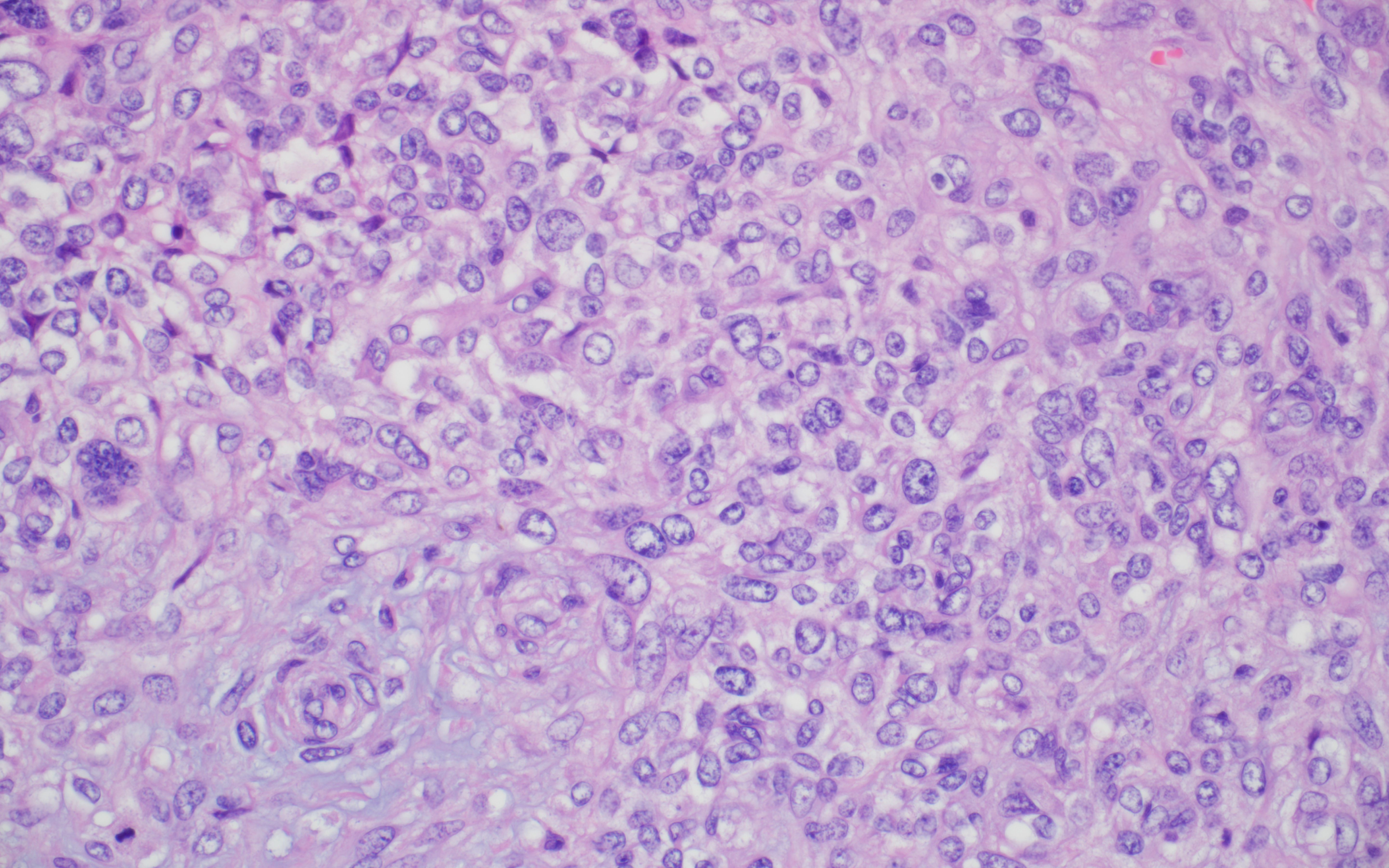 | |
Fig. 5 | |
 | |
Fig. 6 | |
 | ajshfdjskahfkjhdsajkfhkjsdahfjdshfjkdhskfhaskjzhfkashfkasdhfjkashdfkj |